 Manual home
Send Feedback
Manual home
Send Feedback
 Print
Print
|
|
|
|
|
Introduction Oxygenscan
The Oxygenscan measures the relative oxygen pressure at the critical point the RBCs starts to sickle. This so called "point of sickling" (see Figure. 1) quantitatively and reproducibly describes the defined loss of deformability of sickle cell disease (SCD) RBCs.
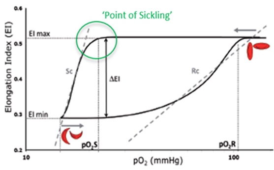
Fig.1 Graphic representation of the measured loss of deformability, the point-of-sickling of SCD RBCs, as a result of oxygen-depletion in time, followed by subsequent gain of deformability of RBC’s during reoxygenation, as is visualized on the Oxygenscan. It is described by pO2: Oxygen- pressure (controlled, in mmHg), and by EI: elongation index (of the RBCs in shear rate), in SC RBCs: Sickle Cell samples Red Blood Cells.
Oxygenscan read out parameters are:
- EImax, which represents RBC deformability at normoxia;
- EImin represents deformability upon deoxygenation;
- The point of sickling (PoS), the point at which a >5% decrease in EI is observed during deoxygenation, reflecting the specific pO2 at which sickling begins (Figure 1).
This PoS is the most characteristic parameters of this measurement. It is typical for each sickle cell RBC sample and sensitive to its status; the PoS shifts due to alteration in treatment.
Supported by the (dynamics of) the change in cell deformability (Elongation index; EI), this measurement of typical RBC behaviour directly reflects the status of the sample susceptibility to oxygen depletion.
The Oxygenscan measurement represents the ‘whole population’ of RBCs (incl. HbS as well as HbF containing cells).
See Also |
Mechatronics home
Send Feedback
Print
|
Page last reviewed: 13/04/2023 14:42:12 (Version: 5.08 (24-04-2023) MRN-231-EN) ©2022 RR Mechatronics |